
Williams Fund research, Oxford University – March 2019 Update from Dr Helen Townley, William Dodd Research Fellow.
Research will work on developing novel nanotherapeutics for childhood cancers. Cancers such as rhabdomyosarcoma, Ewing sarcoma and osteosarcoma have high rates of resistance to conventional chemotherapy, and new approaches are needed. The funding raised by the Williams Fund supports research in the group of Dr Helen Townley which includes DPhil students, MSc students, and undergraduate project students.
Introduction:
Nanoparticles are defined as being smaller than 100 nm. This means that they are approximately 100,000 times smaller than the diameter of a human hair. To see the size and shape of the particles we need to use transmission electron microscopes (TEM) or scanning electron microscopes (SEM). Nanoparticles have a relatively larger surface area when compared to the same volume of material made up of bigger particles. You can see in Figure 1 that the same mass of material can be divided into one large particle, 1 million particles which are 100 times smaller, or 1 billion nanoparticles which are 1000 times smaller. Therefore by simply breaking the particle into smaller units the surface area increases by over 1000x.

Figure 1: Illustration from Buzea et al., 2007
The particles shown below are silica nanoparticles that we have made in the lab (Figure 2). They are approximately 100 nm in diameter, and the pores (a) and channels (c) are approximately 2 nm in diameter.
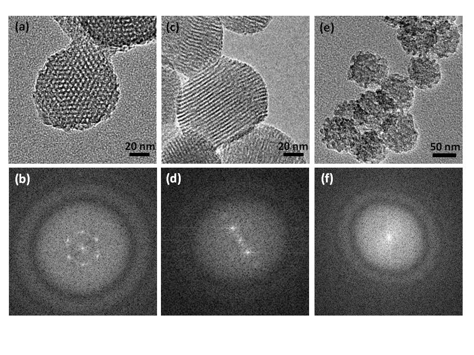
Figure 2: Porous silica nanoparticles
Nanoparticles have such an extremely high surface area & because of the nanometre sized pores so they are able to carry a large drug payload to the tumour.
In addition to be an effective treatment, one of the aims is also to minimize side effects. If the nanoparticles carrying the anti-cancer agent can be concentrated in the tumour then a much higher dose can be delivered directly to cancerous cells. If there was no targeted delivery then the amount of drug could be too high to give to a patient since it would be too damaging to healthy tissues. However, a very high dose directly at the tumour site will give a much better outcome. Targetting can be achieved using nanoparticles because of their very small size. When a tumour reaches a critical size (about 2 mm) diffusion is no longer sufficient to supply the tissue with oxygen and nutrients. The tumour then sends out signals to begin the process of angiogenesis, or vein growth. However, due to the rapid growth of the new veins, they are abnormal and have increased leakiness. The leakiness is due to gaps in the vein wall called fenestrations, which can be up to 350 nm in size. This means that the nanoparticles in the blood circulatory system which are smaller than 350nm can leave the blood stream and accumulate in the tumour. Since the tumours also don’t have a proper lymph system, the nanoparticles are retained in the tumour and so can deliver the drug over a period of time, or be activated repeatedly. Nanoparticles can also be targeted to tumours using antibodies. The antibodies can be designed to recognize proteins that are only expressed on the surface of cancerous cells. It is much easier to add antibody targetting to a nanoparticle than to a small molecules drug. This is because the antibody does not directly interact with the drug and so won’t change its active properties.
Nanoparticles can be made from many different materials, and in addition to silica we have used e.g. doped titanium dioxide to enhance radiotherapy, and protein to encapsulate drugs. We have recently published papers in peer reviewed journals. One piece of research looked at the ability of Citral to kill cancer cells. The compound was effective at destroying rhabdomyosarcoma cells but it is very volatile and breaks down quickly into inactive substances. We encapsulated the compound in protein nanoparticles which stabilized the compound. We then embedded the nanoparticles in a small bio-degradable wafer. This was designed so that after surgical removal of the tumour the wafer could be left in the empty tumour bed, and release the anti-cancer agent over several weeks. This could be important since there is often recurrence of a tumour at the site of removal, due to a small number of cancer cells being left behind. The full paper can be accessed by clicking here.
Another recent publication looked at whether we could make a biocompatible nanoparticle to treat tumours by starving them of iron. Cancer cells often have different iron metabolism than healthy cells. One reason is that cancer cells grow very rapidly and therefore need lots of iron. There are a number of different compounds which can bind, or chelate, iron. However, you cannot simply treat the whole body with chelators because healthy cells also need iron to grow. We therefore synthesized nanoparticles from melanin. This is a naturally occurring pigment in the body which in addition to other uses, is known to bind iron. The nanoparticles that we synthesized were around 220 nm and were effective at binding iron in solution. We were also able to show that they effectively killed cancer cells without harming healthy cells. Also, because they are nanoparticles they can be targeted to the tumour and not deplete the rest of the body of iron. Read the full publication here.
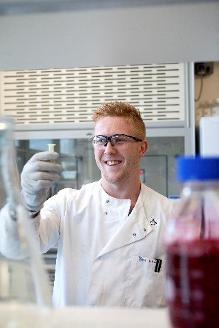
Ben White
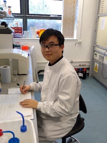
Chengchen Duan
Ben White and Chengchen Duan started their research in nanoparticle therapies for paediatric cancer in October 2018. Ben decided that after completing his undergraduate degree at Bath University that he would like to return to my group to study for a DPhil degree. (He spent a placement year working in Oxford). Ben is self-funded and received a rugby scholarship from St Edmund’s college. However, since there was no support for his research, the project would not have been possible without Williams Fund support. Chengchen received a China-UK scholarship to support his personal and university tuition, however, he does not have funding to cover his laboratory work. Without support from the Williams Fund it would not be possible to undertake this work.
While the core themes of our research remain the same, new research will put more emphasis on remote activation of the nanoparticulate systems for release only at the tumour site. We are currently developing gold and silica nanoparticles with pH responsive coatings. Since the pH within a tumour is lower than surrounding tissues due to metabolic differences, the anticancer agent would only be released at the tumour location. We are also preparing iron oxide nanoparticles with a polymer coating which will permit actuation with magnetic radiation which will result in a temperature increase and alter the polymer coating. Furthermore, targeting elements are being incorporated such as aptamers. Aptamers can be used in a similar way to antibodies, but they are cheaper to produce, and are less likely to elicit an immune response in the body.
Other projects with MSc students and undergraduates will also include testing novel compounds from plants which have potential anticancer activities, but due to their pharmacokinetics would benefit from nanoparticle encapsulation and comparing the effects on paediatric or adult glioblastoma cell lines.
Dr Helen Townley, William Dodd research fellow.
To download this article click here: Research update from HT March 2019